Upper Canada Animal Hospital

Elbow and Hip Dysplasia

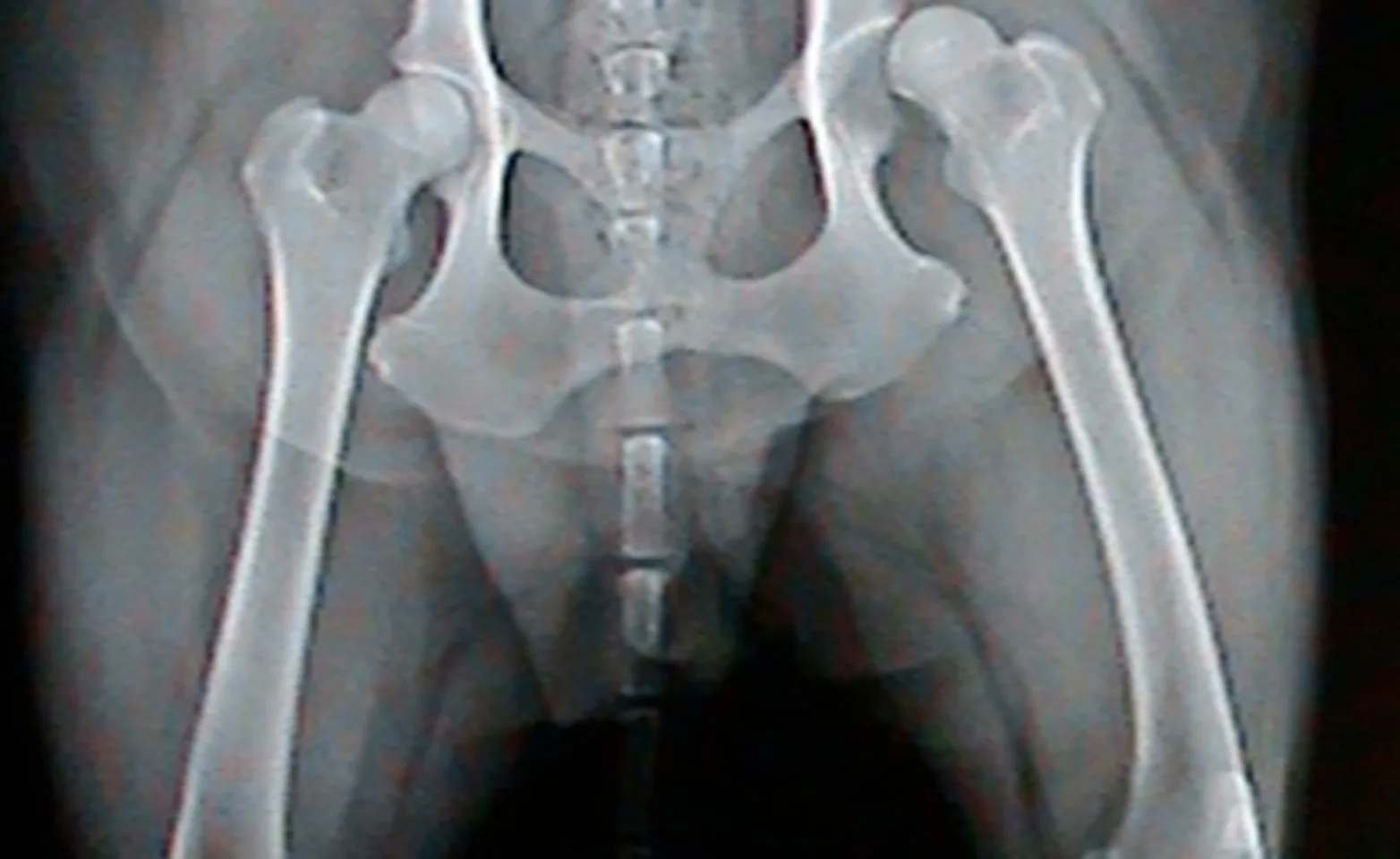
At Upper Canada Animal Hospital we have seen innumerable cases of hip dysplasia or arthritis in dogs and cats. With this condition, the hip joint develops abnormally such that the bones within the joint do not form a secure, tight fit. It’s an extremely painful condition for pets and often results in severe wear and tear of the hip joint.
Hip dysplasia is a condition affecting both dogs and cats. With this condition, the hip joint develops abnormally such that the bones within the joint do not form a nice tight fit. The wear and tear that results from hip dysplasia lead to a flattened femoral head (top of the thigh bone that sits in the hip joint) that gradually slips out of a progressively more flattened joint socket. New bone may develop in the area, causing arthritic pain—this and the laxity (looseness) in the joint may lead to lameness and reduced function.
There are basically two categories of hip surgery when it comes to hip dysplasia. These differ in that one involves early surgical intervention as a preventive measure while the other focuses on alleviating the pain and lameness associated with an unstable arthritic hip joint. This involves surgery on immature dogs in an attempt to improve hip stability so as to minimize future arthritis and pain. The second focuses on treatment of a painful arthritic hip joint. Unfortunately the vast majority of hip surgeries are performed on mature dogs that have developed severe arthritis, are painful, and have not responded adequately to medical management since early detection of hip dysplasia can be quite difficult and require specific tests.
Elbow and Hip Dysplasia FAQ
How do you, as a pet owner, check your pet for hip dysplasia or arthritis?
Pets with hip dysplasia will typically appear uncomfortable and stiff
Often, they exhibit reluctance in running, climbing stairs, or getting into the car
You should be concerned if they have trouble sitting down or getting up
Overall decreased activity
In some cases, your pet may even show loss of muscle mass in the hind legs
Your pet may develop an unsteady gait
“Bunny Hop” when running
Enlargement of shoulder muscles – placing weight on forelimbs
At the first sign of trouble, get your cat or dog to a professional animal hospital – the earlier the diagnosis, the faster the recovery. Upper Canada Animal Hospital is well equipped with the expertise and radiographic equipment required to diagnose hip dysplasia and the skills to treat it in a timely manner.
What Causes Hip Dysplasia/ Arthritis in Animals?
The two most important factors to consider when discussing hip dysplasia are environmental and genetic:
Genetic susceptibility to hip looseness or laxity
Rapid weight gain and obesity
Pelvic-muscle mass
Nutritional factors
Trauma
Can Hip Dysplasia be prevented?
As the old saying goes “an ounce of prevention is worth a pound of treatment”. Prevention starts by selecting a breeder that has made a genuine effort to minimize the likelihood of the condition developing in the dogs they produce. In breeds that are prone to hip dysplasia, it is imperative that the breeding dogs have been certified free of the condition (no excuses that a breeder may offer are acceptable) as this condition has a genetic component and is therefore passed down from the parents to their offspring. It is also very important that puppies be fed well-balanced diets and are kept in lean body condition and in good muscle mass as this will also reduce the strain on the hip joints during their development.
Is surgery always necessary?
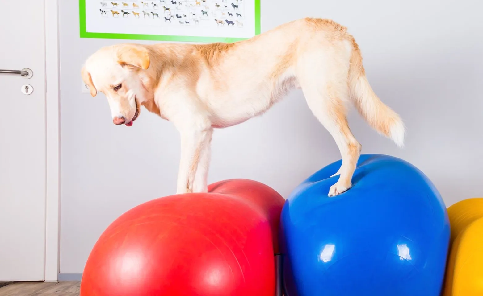
Thankfully no. Even in severe cases of hip dysplasia, surgery can often times be avoided if the pet is kept lean and in good muscle mass. Medications, laser therapy, physiotherapy, and supplements can also be of tremendous benefit as they can further decrease or even eliminate the pain associated with hip dysplasia. Young dogs (often between 4 and 18 months) may show severe signs of hip discomfort/pain then gradually improve over the course of months. For this reason, Dr. Turpel generally recommends against surgery in these young dogs unless medical management has been largely unsuccessful.
When is surgery indicated?
Surgery is typically indicated for pets that do not respond to medical management. Medical management is a large umbrella that covers many treatments. These include but are not limited to: anti-inflammatories, weight loss, laser therapy, physiotherapy, supplements, diet, acupuncture, etc. Dr. Turpel defines successful medical therapy as the pet is kept comfortable, has an excellent quality of life and is able to perform “normal” activities (walking, running, playing) in a pain-free fashion.
What surgeries are available?
If surgery is deemed necessary there are two main surgical options for hip dysplasia or for any causes of severe arthritis associated with the hip joint(s).
The first procedure is a Total Hip Replacement (THR). This has been considered the gold standard of hip surgery over the last 2 to 3 decades. Since its inception, numerous improvements in both the implant materials and surgical technique now result in claims of 70% to 90% success rates for return to excellent pain-free function of the leg(s). The surgery involves the creation of an artificial ball and socket joint by removing the natural components (head of the femur- “ball”, and acetabulum of the pelvis- “socket”) of the hip joint and inserting synthetic replacement parts that result in a smooth, stable, pain-free hip joint(s). Unlike people, the lifespan of our pets is such that a one-time prosthesis should do, and repeat surgery to replace worn implants is not typical. After surgery, one can expect a pet to use the limb well in a week or two, but exercise restriction usually lasts for 2-3 months. Physiotherapy is typically recommended during recovery to assist a return to mobility, and for maintenance of muscle strength and joint flexibility. Despite the advancements in the THR surgery a 10% to 30% complication rate can still be expected. Unfortunately, many of these complications can be extremely serious since they may involve the fracturing of bones, breakage or loosening of implants, dislocation of the artificial hip and severe infection. Despite the excellent outcome in the majority of cases these complications in conjunction with the cost of the procedure often times limit its application.
The second surgical procedure is a Femoral Head and Neck Ostectomy (FHO). The FHO involves the removal of the entire head and neck of the femur thereby removing the pain associated with the grinding within the hip joint. Once these structures are removed a fibrous pad develops in the area between the femur and pelvis and results in stability of the hip joint and a pain-free condition. This procedure is performed quite frequently since it typically results in a good return to pain-free function of the limb(s), has a low complication rate, and is considered far more affordable than a THR. However, patient selection is far more important in FHO’s than in THR’s since the FHO is not initially as stable as the THR and the surgery has a much longer recovery period ( good weight bearing seem typically within 1 month and full recovery in 4 to 6 months). It is also generally accepted that the success of the FHO decreases with the increased weight of the patient.
Many surgeons are reluctant to perform the surgery on large dogs while others, Dr. Turpel included, report good success in patients up to 45 kilograms if they are otherwise healthy and not too overweight. FHO’s are considered a salvage procedure since once completed they cannot be reversed or other procedures (i.e. THR) performed. FHO’s are frequently performed if serious complications with a THR are encountered.
The Triple Pelvic Osteotomy (TPO) is the third procedure for hip dysplasia and is briefly mentioned since it is still occasionally performed, however, it has largely fallen out of favour by many surgeons over the last twenty years as the necessity and success of the procedure has been questioned. This procedure involves realigning the bones of the hip joint in young dogs, prior to the development of arthritis, so as to increase the stability of the hip joint in hopes of preventing or minimizing future arthritis and pain.
If you suspect that your pet suffers from hip disease please contact your veterinarian since very successful medical and surgical options are available for this very painful condition.

Elbow Dysplasia in Dogs
Elbow dysplasia is a complex, inherited disease which primarily affects intermediate and large breed dogs however this condition may be seen in any breed. Breeds that appear to be particularly predisposed to elbow dysplasia include Bernese Mountain Dog, German Shepherd, Rottweiler, Golden Retriever, Labrador Retriever, Newfoundland, Saint Bernard, Mastiff, Springer Spaniel, Australian Shepherd, Chow Chow, Shar-Pei, Shetland Sheepdog, and some Terrier breeds.
While both elbows are frequently affected the condition can present unilaterally. Elbow dysplasia is characterized by varying degrees of elbow incongruity, bony fragments (bone chips), and ultimately, severe arthritic changes. Rather than being a specific diagnosis, elbow dysplasia encompasses a number of developmental abnormalities. These abnormalities typically develop between four and eight months of age and include osteochondritis dissecans (OCD) of the medial humeral condyle, ununited anconeal process (UAP), joint incongruency, and fragmentation of the medial coronoid process (FCP). A dog with elbow dysplasia may have one or more of the aforementioned abnormalities. Regardless of the specific condition, the best outcomes are obtained with early diagnosis and prompt treatment. Minimally invasive treatment modalities should be used whenever possible. Long-term medical management of osteoarthritis may also be needed as adjunctive therapy since arthritis will often times progress regardless of treatments employed.
Dogs affected with elbow dysplasia will have symptoms that range from mild, intermittent lameness to that of severe and crippling. The gait is often characterized by excessive paddling or flipping of the front feet. The dog may either hold the elbows out or tucked in and often stands with the feet rotated outward. Affected dogs may also sit or lie down much of the time or play for shorter periods with other dogs of comparable age. They are often described as quiet or even lazy. Frequently, they are stiff when rising, have difficulty or are slow going downstairs, and tire easily. Exercise typically makes the lameness worse. In dogs with bilateral elbow dysplasia, the lameness may seem intermittent or shift from one front leg to the other. When both front legs hurt, dogs do not limp constantly. Rather, they shift weight from their elbows by altering their gait and stance. These dogs will only limp when one elbow is more painful than the other. On examination, manipulation of the affected elbow(s) is often resisted (due to discomfort). Swelling and crepitus (grating) may be palpated with the swelling worse after exercise. In some cases, the joint will be thickened. Muscle atrophy may also be present due to disuse of the leg and range of motion of the elbow may be severely restricted in advanced cases.

Treatment options for elbow dysplasia
Osteochondrosis of medial humeral condyl: removal of OCD flap and abrasion arthroplasty or microfracture of the subchondral bone. (Preferably performed with the aid of an arthroscope to gain better visualization of the cartilage lesion.)
Ununited Anconeal Process:
If the Anconeal process appears healthy and non-displaced a proximal dynamic ulnar osteotomy +\- lag screwing of the anconeal process is recommended.
If the Anconeal process appears healthy but is displaced or mobile, but otherwise appears healthy a proximal dynamic ulnar osteotomy combined with lag screwing of the process is considered to be the best option.
If the anconeal process appears unhealthy, arthritic changes or fragmented, it should be removed.
In all cases of ununited anconeal processes, the medial coronoid should be evaluated since approximately 20% of dogs with UAP will also have FCPs.
Elbow incongruency: If the ulna is shorter than the radius a dynamic proximal ulnar osteotomy is indicated.
Fragmented Medial Coronoid: This is by far the most controversial aspect of elbow dysplasia. It is generally accepted that even with surgical intervention all dogs will experience progression of degenerative joint disease (arthritis). The goal of surgery in these dogs is to delay or minimize the progression of arthritis while decreasing a patients discomfort. Treatments range from medical treatments (I will outline these below since they are similar for all cases of elbow dysplasia) to removing the fragment(s) and cleaning up the adjacent bone, through to procedures that realign the elbow joint to decrease strain/pressure on the abnormal medial compartment of the joint (with FCPS the medial compartment of the joint collapse resulting in further strain pressure and pain in the elbow).
Medical management is used to provide pain relief but has not been shown to be very effective in the vast majority of cases, and as arthritis progresses this management becomes less and less effective. This option is typically used if the elbow already has significant arthritis that may not respond as well to surgery as a “clean” elbow would, and if cost is a major limiting factor in proceeding with surgery.
Fragment removal has been the cornerstone of treatment of FCP for a number of years. Many dogs improve, especially in the short- term to this surgery, however as arthritis and collapse of the medial compartment progresses many of these dogs will become clinically lame again. This deterioration in time occurs due to the progression of arthritis and collapse of the medial joint compartment- both resulting in increased discomfort. This procedure is considered superior to medical management alone however for the vast majority of dogs this will not provide excellent long-term results. (Preferably performed with the aid of an arthroscope to gain better visualization of the fragment and surrounding bone).
PAUL Procedure: Due to the progression of arthritis and lameness typically seen in dogs with medical management and fragment removal the PAUL (Proximal Abducting Ulnar Osteotomy) procedure has been developed. This procedure has been designed to realign the elbow joint so as to take the weight off the painful, severely damaged and often times collapsed medial compartment. The PAUL has been tested and clinically evaluated in Europe and has shown impressive results in the majority of dogs treated. It is very important to note that many of the dogs treated already exhibited signs of moderate arthritis in their elbows.

Weight loss: These dogs need to be thin. An exercise program that takes into account their exercise abilities combined with dietary management is necessary ( see your veterinarian for diet foods) and recommendations for low impact activities.
Rehab: Get the elbow mobile and rebuild lost muscle mass.
Anti-inflammatories or other pain medications: These are needed to provide comfort and to help increase exercise levels that would otherwise be too uncomfortable.
Supplements: Omega 3 fatty acids, glucosamine, chondroitin etc. ( be aware the only good quality studies, that I am aware of at the current time, that show any evidence to benefits of supplements involves high-quality Omega 3 fatty acids).
Chondroprotectants: Cartrophens, Adequan – have been thoroughly studied and shown to be beneficial in approximately 80% of cases.
Avoid high impact activities that affect the elbow. Going down the stairs, jumping down, ball chasing etc.- these activities put tremendous strain on the elbows)
